|
||||||||||||||||||||||||||||||||||||||||||||||||||||||
Cervical cancer http://www.cancer.gov/cervix
Cervical cancer Background Cancer of the cervix is now thought to be associated by certain strains of a sexually transmitted virus (human papilloma virus). The role of other sexually transmitted infections is unclear. To date no association has been found between invasive cancer of the cervix and HIV infection. Other related risk factors include early age at first intercourse, number of sexual partners, parity and poor socio-economic conditions. Cancer of the cervix is the most common cancer in women in developing countries. Rates for this cancer have been declining in developed countries, partly as a result of improved socio-economic circumstances, better access to medical facilities and due to screening. A national screening programme using a Papanicolaou (Pap) smear, three times in a woman lifetime (at about 35, 45 and 55 years) and with an 80% coverage will reduce the incidence of this cancer by half in the screened women.
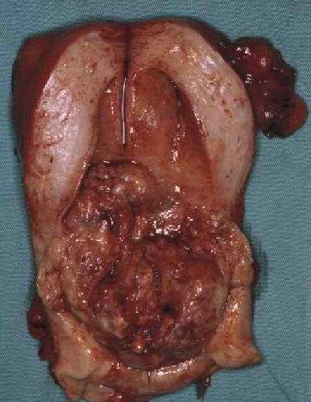
NUMBERS & INCIDENCE Between 1993 and 1995, an average of 3387 new cases of cancer of the cervix were reported. By contrast, 1,497 deaths from cancer of the cervix were reported for 1994 by the CSS. The crude incidence rate was 17/100 000 and the ASIR 22/100 000. Cancer of the cervix is the most common cancer in women (Lifetime Risk = 1 in 41). Important population differences exist: in black females the LR was 1 in 34, whereas in whites 1 in 93, about a threefold difference. In Asian and coloured females the LR was about 1 in 50. There appears to be a reduction in the incidence of cancer of the cervix in females from 1:30 in 1992 to 1:40 in the subsequent three years. The relative frequency of this condition has however also dropped from 17.9 of all cancers to 16.7% and the relative risk between black and white from 3.3 to 2.7. A more formal trends analysis is required to determine whether or not there is a real decline in cervical cancer over the period, but for the first time, breast cancer has overtaken cervical cancer. Cervical cancer is a disease in which malignant (cancer) cells form in the tissues of the cervix. Human papillomavirus (HPV) infection is the major risk factor for development of cervical cancer. There are usually no noticeable signs of early cervical cancer but it can be detected early with yearly check-ups. Possible signs of cervical cancer include vaginal bleeding and pelvic pain. Tests that examine the cervix are used to detect (find) and diagnose cervical cancer. Certain factors affect treatment options and prognosis (chance of recovery). Cervical cancer is a disease in which malignant (cancer) cells form in the tissues of the cervix.The cervix is the lower, narrow end of the uterus (the hollow, pear-shaped organ where a fetus grows). The cervix leads from the uterus to the vagina (birth canal). Cervical cancer usually develops slowly over time. Before cancer appears in the cervix, the cells of the cervix go through changes known as dysplasia, in which cells that are not normal begin to appear in the cervical tissue. Later, cancer cells start to grow and spread more deeply into the cervix and to surrounding areas. Human papillomavirus (HPV) infection is the major risk factor for development of cervical cancer. Infection of the cervix with human papillomavirus (HPV) is the most common cause of cervical cancer. Not all women with HPV infection, however, will develop cervical cancer. Women who do not regularly have a Pap smear to detect HPV or abnormal cells in the cervix are at increased risk of cervical cancer. Other possible risk factors include the following: Giving birth to many children. Having many sexual partners. Having first sexual intercourse at a young age. Smoking cigarettes. A diet lacking in vitamins A and C. Oral contraceptive use ("the Pill"). Weakened immune system. There are usually no noticeable signs of early cervical cancer but it can be detected early with yearly check-ups. Early cervical cancer may not cause noticeable signs or symptoms. Women should have yearly check-ups, including a Pap smear to check for abnormal cells in the cervix. The prognosis (chance of recovery) is better when the cancer is found early. Possible signs of cervical cancer include vaginal bleeding and pelvic pain. These and other symptoms may be caused by cervical cancer or by other conditions. A doctor should be consulted if any of the following problems occur: Vaginal bleeding. Unusual vaginal discharge. Pelvic pain. Pain during sexual intercourse. Tests that examine the cervix are used to detect (find) and diagnose cervical cancer. The following procedures may be used: Pap smear: A piece of cotton, a brush, or a small wooden stick is used to collect cells from the cervix and vagina. The cells are viewed under a microscope. Abnormal (precancerous) cells in the tissues of the cervix can usually be found by a Pap smear. Colposcopy: The tissues of the vagina and cervix are examined using a lighted magnifying instrument called a colposcope. Biopsy: If abnormal cells are found in a Pap smear, the doctor may do a biopsy. A sample of tissue is cut from the cervix and viewed under a microscope. A biopsy that removes only a small amount of tissue is usually done in the doctor's office. A woman may need to go to a hospital for a cervical cone biopsy (removal of a larger, cone-shaped sample of cervical tissue). Pelvic exam: A procedure to check the uterus, vagina, ovaries, fallopian tubes, bladder, and rectum to find any abnormality in their shape or size. Endocervical curettage: A curette (a spoon-shaped instrument) is used to collect cells from the cervical canal. The cells are viewed under a microscope. This procedure is sometimes done at the same time as the colposcopy. Certain factors affect treatment options and prognosis (chance of recovery). The treatment options and prognosis (chance of recovery) depend on the stage of the cancer (whether it affects part of the cervix, involves the whole cervix, or has spread to the lymph nodes or other places in the body), the type of cervical cancer, the size of the tumor, and the patient's desire to have children. Lymph nodes are small, bean-shaped structures found throughout the body. They filter substances in a fluid called lymph and help fight infection and disease. Treatment of cervical cancer during pregnancy depends on the stage of the cancer and the stage of the pregnancy. For cervical cancer found early or for cancer found during the last trimester of pregnancy, treatment may be delayed until after the baby is born.
Stages of Cervical Cancer
* The depth of incvasion should not be more than 5 mm taken from the base of the epithelium, either surface or glandular, from which it originate. Vascular space involveme,ent, either venous or lymphatic, should not alter the staging Notes Diagnosis of both Stages IA1 and IA2 is based on microscopic examination of removed tissue, preferably a cone, which must include the entire lesion. The lower limit of Stage IA2 should be measurable macroscopically (even if dots need to be placed on the slide prior to measurement). The upper limit of IA2 is determined by measurement of the two largest dimensions in any given section. Revised staging - adopted in 1988 - varies from the previous staging primarily in the stage I category. These changes have occasioned a good deal of controversy and a substantial body of opposition from some gynecologic oncologists. The defined limits of the 1a2 often appear impractical in clinical practice. Multiple foci of invasion may be present, the cone biopsy may not include the entire lesion, and prior colposcopic biopsies which encroach on the lesion may alter the volumetric dimensions. A major concern is that clinicians will interpret the stage Ia2 lesion as one that can be approached in a more conservative manner such as simple hysterectomy. Taken as a group, a retrospective study done here suggested that such lesions carry a risk of nodal metastases in excess of 4% and a recurrence rate of 6% even when treated by radical hysterectomy. The G.O.G. previously had described "microinvasion" as a lesion with less than 3 mm of invasion, no vascular space involvement and no areas of confluence. as possibly suitable for conservative treatment. It has been suggested that this guideline be substituted in treatment planning. Cervical cancer remains the one remaining major gynecologic cancer that is subjected to "clinical" staging as opposed to surgical staging. This decision, of course, reflects an appreciation that a large number of cases - perhaps a majority - are treated with radiation therapy without surgical intervention. It is generally agreed that the most experienced clinician involved in the case should stage the lesion and if there is a doubt as to which stage applies, the earlier stage is mandatory. Examination for clinical staging permits palpation, inspection, colposcopy, hysteroscopy, curettage, cystoscopy, proctoscopy and roentgen examinations to include X-rays of the lungs and skeleton and intravenous urography. Other procedures such as laparoscopy or lymphography may not be employed. A conization of the cervix is regarded as a clinical examination. An IVP revealing hydronephrosis or a nonfunctioning kidney attributable to stenosis of the ureter by cancer permits the allotment of a case to Stage III regardless of other factors. After cervical cancer has been diagnosed, tests are done to find out if cancer cells have spread within the cervix or to other parts of the body. The process used to find out if cancer has spread within the cervix or to other parts of the body is called staging. The information gathered from the staging process determines the stage of the disease. It is important to know the stage in order to plan the best treatment. The following tests and procedures may be used in the staging process: Chest x-ray: Brief exposure of the chest to radiation to produce an image of the chest and its internal structures. CT scan (CAT scan): A CT scan creates a series of detailed pictures of areas inside the body, taken from different angles. The pictures are created by a computer linked to an x-ray machine. This test is also called computed tomography, computerized tomography, or computerized axial tomography. Lymphangiography: An x-ray is made of the lymph system. A dye is injected into a lymph vessel and travels through the lymph system. The dye outlines the lymph vessels and organs on the x-ray. This test helps determine whether cancer has spread to the lymph nodes. Pretreatment surgical staging: Surgery (an operation) is done to find out if the cancer has spread within the cervix or to other parts of the body. In some cases, the cervical cancer can be removed at the same time. Pretreatment surgical staging is usually done only as part of a clinical trial. Ultrasound: A test that uses sound waves to create images of body tissues. MRI (magnetic resonance imaging): A procedure in which a magnet linked to a computer is used to create detailed pictures of areas inside the body. This test is also called nuclear magnetic resonance imaging (NMRI). The results of these tests are viewed together with the results of the original tumor biopsy to determine the cervical cancer stage. Recurrent Cervical Cancer Recurrent cervical cancer is cancer that has recurred (come back) after it has been treated. Recurrent cervical cancer may come back in the cervix or in other parts of the body. Treatment Option Overview Key Points for This Section There are different types of treatment for patients with cervical cancer. Three types of standard treatment are used: Surgery Radiation therapy Chemotherapy Other types of treatment are being tested in clinical trials. There are different types of treatment for patients with cervical cancer. Different types of treatment are available for patients with cervical cancer. Some treatments are standard (the currently used treatment), and some are being tested in clinical trials. Before starting treatment, patients may want to think about taking part in a clinical trial. A treatment clinical trial is a research study meant to help improve current treatments or obtain information on new treatments for patients with cancer. When clinical trials show that a new treatment is better than the "standard" treatment, the new treatment may become the standard treatment. Clinical trials are taking place in many parts of the country. Information about ongoing clinical trials is available from the NCI Cancer.gov Web site. Choosing the most appropriate cancer treatment is a decision that ideally involves the patient, family, and health care team. Three types of standard treatment are used: Surgery Surgery (removing the cancer in an operation) is sometimes used to treat cervical cancer. The following surgical procedures may be used: Conization: Surgery to remove a cone-shaped piece of tissue from the cervix and cervical canal for biopsy. Also called cone biopsy. Hysterectomy: The uterus and cervix are removed in a hysterectomy. If the uterus is taken out through the vagina, the operation is called a vaginal hysterectomy. If the uterus is taken out through an incision (cut) in the abdomen, the operation is called a total abdominal hysterectomy. Bilateral salpingo-oophorectomy: The removal of both ovaries and both fallopian tubes. Radical hysterectomy: This surgery involves removing the cervix, uterus, fallopian tubes, ovaries, and part of the vagina. Lymph nodes may also be removed. Pelvic exenteration: If the cancer has spread throughout the pelvis, then the lower colon, rectum, or bladder (depending on where the cancer has spread) may be removed along with the cervix, uterus, and vagina. Plastic surgery may be needed to make an artificial vagina after this operation. Cryosurgery: An instrument is used to freeze and destroy the abnormal tissue. This procedure is also called cryotherapy. Carcinoma in situ may be treated with cryosurgery. Laser surgery : A laser beam (a narrow beam of intense light) is used as a knife to remove the cancer. A laser beam can also be used to kill the cancer cells. This may be called laser therapy. Loop electrosurgical excision procedure (LEEP): An electrical current passed through a thin wire loop is used as a knife to remove abnormal tissue. Radiation therapy Radiation therapy is the use of x-rays or other types of radiation to kill cancer cells and shrink tumors. Radiation therapy may use external radiation (using a machine outside the body) or internal radiation. Internal radiation involves putting radioisotopes (materials that produce radiation) through thin plastic tubes into the area where cancer cells are found. Both external and internal radiation are used for cervical cancer. Chemotherapy Chemotherapy is the use of drugs to kill cancer cells. Chemotherapy may be taken by mouth, or it may be put into the body by inserting a needle into a vein or muscle. Either type of chemotherapy is called systemic treatment because the drugs enter the bloodstream, travel through the body, and can kill cancer cells throughout the body. Other types of treatment are being tested in clinical trials. TREATMENT OPTION OVERVIEWHow cancer of the cervix is treated There are treatments for all patients with cancer of the cervix. Three kinds of treatment are used:
A doctor may use one of several types of surgery for carcinoma in situ to destroy the cancerous tissue:
A doctor may remove the cancer using one of these operations:
Radiation therapy is the use of x-rays or other high-energy rays to kill cancer cells and shrink tumors. Radiation may come from a machine outside the body (external radiation) or from putting materials that produce radiation (radioisotopes) through thin plastic tubes into the area where the cancer cells are found (internal radiation). Radiation may be used alone or in addition to surgery. Chemotherapy is the use of drugs to kill cancer cells. Chemotherapy may be taken by pill, or it may be put into the body by a needle inserted into a vein. Chemotherapy is called a systemic treatment because the drugs enter the bloodstream, travel through the body, and can kill cancer cells outside the cervix. Treatments for cancer of the cervix depend on the stage of the disease, the size of the tumor, and the patient's age, overall condition, and desire to have children. Treatment of cervical cancer during pregnancy may be delayed depending on the stage of the cancer and how many months a patient has been pregnant. Standard treatment may be considered because of its effectiveness in patients in past studies, or participation in a clinical trial may be considered. Not all patients are cured with standard therapy and some standard treatments may have more side effects than are desired. For these reasons, clinical trials are designed to find better ways to treat cancer patients and are based on the most up-to-date information. Clinical trials are ongoing in most parts of the country for most stages of cancer of the cervix. To learn more about clinical trials, call the Cancer Information Service at 1-800-4-CANCER (1-800-422-6237); TTY at 1-800-332-8615. STAGE 0 CERVICAL CANCERStage 0 cervical cancer is sometimes called carcinoma in situ. Treatment may be one of the following:
STAGE I CERVICAL CANCERTreatment may be one of the following depending on how deep the tumor cells have invaded into the normal tissue:
STAGE II CERVICAL CANCERTreatment may be one of the following:
STAGE III CERVICAL CANCERTreatment may be one of the following:
STAGE IV CERVICAL CANCERTreatment may be one of the following:
RECURRENT CERVICAL CANCERIf the cancer has come back (recurred) in the pelvis, treatment may be one of the following:
If the cancer has come back outside of the pelvis, a patient may choose to go into a clinical trial of systemic chemotherapy. Treatment of recurrent cervical cancer may include the following: Pelvic exenteration followed by radiation therapy combined with chemotherapy. Chemotherapy as palliative therapy to relieve symptoms caused by the cancer and improve quality of life. Clinical trials of new anticancer drugs or drug combinations. This summary section refers to specific treatments under study in clinical trials, but it may not mention every new treatment being studied. Information about ongoing clinical trials is available from the NCI Cancer.gov Web site. |
||||||||||||||||||||||||||||||||||||||||||||||||||||||
Copyright © 2010 - All Rights Reserved - 曾志仁醫師