Dr. CJ Tseng

                    曾志仁醫師的網頁  -  衛教園地


Cesarean Section

Cesarean section (C-section) is the delivery of a newborn through a surgical incision in the abdomen and front (anterior) wall of the uterus. The C-section rate may approximate 30% in some hospitals, particularly where high-risk pregnancies are managed. This rate of C-section is usually less for women with uncomplicated pregnancies.

Anatomy and Physiology

  • The uterus or womb is a pear shaped organ that is found in the pelvis at the top of the vagina. The uterus in a woman that is not pregnant does not extend above the pubic bone (Figures 1, 2 and 3)
Figure 1 - Anatomy of the uterus and surrounding organs as seen in a section through the middle of the body. Figure 2 - Uterus as seen from in front.
Figure 3 - View of the uterus, ovaries, Fallopian tubes, and round ligament through a laproscope.
  • In the pregnant woman at the end of pregnancy, the uterus enlarges to approximately 40 centimeters (16 inches) above the pubic bone
  • The cervix is found at the lowermost portion of the uterus and is the opening through which the fetus passes during delivery. Normally, this opening is closed until late in the pregnancy
  • Fertilization occurs within the fallopian tubes. The fallopian tubes are found at the top of the uterus, one on each side. The end of each fallopian tube has fingerlike projections called fimbria which guide the egg from the ovary into the Fallopian tube
  • The fertilized egg then passes from the Fallopian tube into the cavity within the uterus where the fertilized egg implants into the wall of the uterus. The site of implantation becomes the
  • As the egg develops into a fetus the remains attached to the fetus through the umbilical cord. Thus the blood supply to the fetus originates in the uterine wall
  • The umbilical cord extends from the to the fetus where it inserts at umbilicus (belly button) of the fetus

Reasons for the Increasing Rate of C-sections

  • Greater emphasis on "quality survival" for the newborn, not simply survival
  • Continuous fetal monitoring of heart rate increases the number of C-sections for fetal distress
  • More women are waiting to start childbearing until later in life because of their careers. With advancing age of the mother there is a greater risk of having other medical problems, thus the C-section rate increases
  • There has been a decrease in the number of forceps deliveries, which in turn increases the number of C-sections
  • Many obstetricians no longer wish to take the risk of a vaginal breech (bottom first) delivery
  • Women with prior a C-section often choose or are required to have a repeat C-section

Indications for C-section

Some indications are controversial and some are accepted as the standard.

  • Fetal indications
    1. Abnormal fetal heart rate patterns (nonreassuring fetal status) (Figure 4A)
      Figure 4a - Non-reassuring fetal status - Printout showing slowing of fetal heart rate with uterine contractions, which led to cesarean section.
    2. Breech presentation of the fetus ( bottom down instead of head down) (Figure 4B)
      1. Very low birth weight (<1500grams or 3.3 pounds)
      2. Active genital herpes lesions
      3. Idiopathic thrombocytopenia purpura - disease in which there are low platelets in the blood and easy bleeding
      4. Major malformations in the fetus making passage through the birth canal difficult or impossible
        Figure 4b - Breech presentation. Fetus presents bottom side down
  • Maternal indications
    1. Tumor obstructing the birth canal
    2. Large genital warts (condyloma)
    3. Cervical cerclage- a suture is placed in the cervix to prevent preterm delivery. Cerclage may be permanent or temporary to allow for vaginal delivery
    4. Certain vaginal surgeries (vaginal repair can break down)
    5. Conjoined twins (Siamese twins)
    6. Prior surgery in which an incision was made in the uterus (myomectomy -removal of fibroid tumor)
    7. HIV - The American College of Obstetricians and Gynecologists recommends that HIV+ mothers should deliver the fetus by C-section at 38 weeks of pregnancy to reduce the chance of transmitting the virus to the fetus
  • Maternal-Fetal Indications
    1. Cephalopelvic disproportion- either the baby is too large or the pelvis is too small to allow passage
    2. Failure of the cervix to dilate or failure of the fetus to pass down the birth canal
    3. Abruption - when the tears away from the uterus. This is an emergency situation
    4. Previa - this is when the implants over all or part of the cervix. (Figure 4C)
    Figure 4C - previa. The is implanted over the opening of the cervix thus preventing a vaginal delivery.

Making the Diagnosis

  • Electronic fetal monitoring - monitoring of the fetus' heart tracing. Repetitive decreases in heart rate may signal a decrease in oxygen to the fetus (e.g. compression of the umbilical cord)
  • Fetal scalp pH - monitors the acidity of the scalp tissue. An elevated acidic reading (pH < 7.2) indicates a build up of carbon dioxide and lactic acid in the blood, which occurs when the fetus is poorly oxygenated
  • Examination of the cervix -
    1. Rule out prolapse of the umbilical cord
    2. Rule out bleeding from the cervix (e.g. with l previa
  • Abdominal Ultrasound - ultrasound images of the uterus and its contents are obtained
    1. Position of the fetus - head down or up
    2. Position of the relative to the cervix
    3. Size of the head (e.g. enlarged head due to hydrocephalus)
  • Vaginal Ultrasound - similar to abdominal ultrasound but may be more accurate in previa
  • Test for HIV ?MRI - can be used to determine the position of the fetus and
  • Blood tests for Rh factor and bleeding tendency

The Procedure

  • The skin is prepared with a solution that reduces the risk of wound infection
  • A catheter is placed in the bladder
  • The hair near the incision may be shaved
  • An incision is made in the skin and is carried through the abdominal wall to enter the pelvis. The skin incision may be made vertical (up and down) or transverse (from side-to-side). The decision is based on many factors including speed of entry, exposure needed, anticipated weight of the baby and risk of wound infection. A transverse skin incision is most common and is usually made 2-3 centimeters (one inch) above the pubic bone (Figure A)
  • The uterus is then identified. There is a layer of thin tissue, which drapes over the anterior surface of the uterus and then onto the bladder (the vesicouterine peritoneum). This layer is incised so that the bladder can be retracted away form the uterus to allow for the uterine incision. (Figure B) The incision is then carried into the uterus to allow for delivery of the baby
  • The uterine incision is then made down to the amniotic sack (fetal membranes or bag of water). (Figure C)
  • The uterine incision can be either transverse or vertical. Ninety percent have a transverse uterine incision. Some indications for a vertical incision in the uterus are a pre-term fetus, a fetus that is not head down and with emergency C-sections. Even in these situations a transverse incision may sometimes be used. A woman that has a prior C-section with a vertical uterine incision is usually not a candidate for vaginal birth
  • The fetal head or buttocks are then delivered through the uterine incision followed by the rest of the body. (Figure D) Then the is delivered
  • Some obstetricians repair the uterus by first delivering the uterus through the abdominal incision and some repair it while it is still in the abdomen. The uterus is closed with one or two layers of suture (Figure E)
  • The layers of the abdominal wall are sutured and then the skin closed with either suture or staples

Complications

  • Injury to the bladder or ureters
  • Bowel injuries may also occur. Risk factors are prior abdominal surgery, and pelvic or abdominal infections, both of which may lead to adhesions (scar tissue)
  • Uterine atony. A condition in which the uterus does not contract down adequately after delivery and lead to massive hemorrhage. If conservative treatment fails, the surgeon may try to tie off one or both of the arteries to the uterus. A hysterectomy may be required on rare occasions to control the bleeding if other measures fail
  • Wound infection
  • Urinary tract infection
  • Failure of bowel function causing bowel distension
  • Deep venous thrombosis. Blood clots in the veins of the legs or pelvis that may go to the lungs (pulmonary embolus)

Care After Surgery

  • Walking is important to prevent pneumonia and blood clots
  • Diet initiallly consists of ice chips on the day of surgery, fluids on the first day after surgery, and a regular diet two days after surgery
  • The catheter placed in the bladder at the time of surgery,is usually removed the first day after surgery
  • Fluids may be given in the vein up to and sometimes through the first day after surgery
  • Discharge to home may occur in two to five days depending on the circumstances

Vaginal Birth After C-Section (VBAC)

Some women may be a candidate for vaginal birth after Cesarean section

  • Candidates for VBAC are
    1. One or two prior low transverse c-sections
    2. Clinically adequate pelvis (pelvis seems large enough based on exam)
    3. Physician readily available throughout labor capable of performing an emergency c-section.
    4. Available anesthesia and personnel for emergency c-section
  • There is a 60-80% success rate with VBAC. There is a higher success rate when
    1. Prior c-section was performed for abnormal fetal heart tones (nonreassuring fetal status)
    2. The woman has had at least one vaginal birth in the past
  • Complications
    1. Uterine dehiscence at the old incision site. Women who have dehiscence have a partial separation of their old incision and may not need any repair if clinically stable
    2. Uterine rupture at the old incision site. The incidence of rupture is 0.2-1.5%. Women with uterine rupture need an emergency c-section

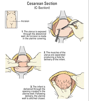
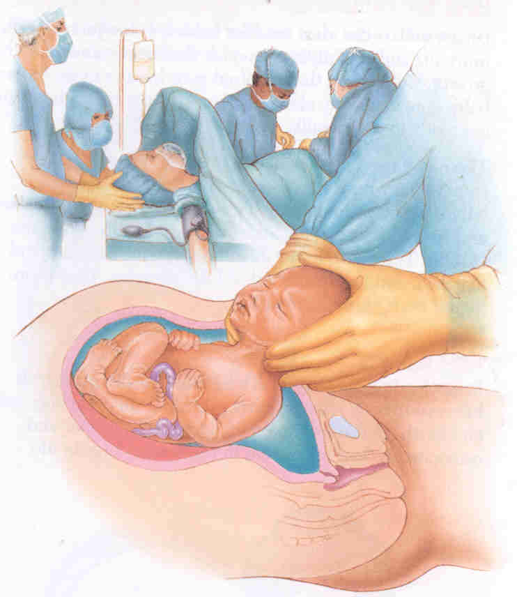
A C-section delivery is performed when a vaginal birth is not possible or is not safe for the mother or child.

Surgery is usually done while the woman is awake but pain-free from the chest to the legs (epidural or spinal anesthesia). An incision is made across the abdomen just above the pubic area. The uterus is opened, the amniotic fluid is drained off, and the baby is delivered. The baby's mouth and nose are cleaned of fluids and the umbilical cord is clamped and cut. The baby is handed to the pediatrician or nurse who will make sure that he is breathing well. The mother is awake and she can hear and see her baby.

Due to increased medicalization of birth, C-sections have become fairly common (up to 20% of all births in the U.S.).


Vertical Inscision

Horizontal or Bikini Inscision


Indications

Some of the main reasons for C-section delivery instead of vaginal delivery include the following

decreased oxygen level

multiple babies in the uterus (multiple births)

increased or decreased heart rate

problems with the cervix

problems with the uterus

too loose or relaxed (incompetent cervix)

too small, scarred, or deformed

active genital herpes infection of the mother

problems with the placenta and umbilical cord

abnormally developed baby

attached too close to the cervix (placenta previa)

"buttocks-first" delivery (breech presentation)

baby's head is too large to pass through mother's pelvis (cephalopelvic disproportion)

crosswise (transverse) position

prolonged labor

pregnancy at older age (over 40 years

abnormal position of the baby

extreme illness (toxemia, preeclampsia, eclampsia, hypertension)

incompletely attached to the uterus (placenta abruptio)

 Convalescence:

The average hospital stay is 2 to 4 days. Recovery takes longer than it would from a natural birth. Walking is encouraged the day of surgery to speed recovery. Pain can be managed with oral medications.

 Risks

reactions to medications

infection of the bladder or uterus

problems breathing

infection of the uterus

bleeding

back pain


PROBLEMS DURING SURGERY

BLEEDING IS NOT CONTROLLED

• Massage the uterus.

• If the uterus is atonic, continue to infuse oxytocin and give ergometrine 0.2 mg IM and prostaglandins, if available. These drugs can be given together or sequentially (Table S-8).

• Transfuse as necessary.

• Have an assistant press fingers over the aorta to reduce the bleeding until the source of bleeding can be found and stopped. 

• If bleeding is not controlled, perform uterine and utero-ovarian artery ligation  or hysterectomy.


BABY IS BREECH

• If the baby is breech, grasp a foot and deliver it through the incision. 

• Complete the delivery as in a vaginal breech delivery:

- Deliver the legs and the body up to the shoulders, then deliver the arms;

- Flex (bend) the head using the Mauriceau Smellie Veit manoeuvre.

BABY IS TRANSVERSE

THE BABY’S BACK IS UP

• If the back is up (near the top of the uterus), reach into the uterus and find the baby’s ankles. 

• Grasp the ankles and pull gently through the incision to deliver the legs and complete the delivery as for a breech baby.

THE BABY’S BACK IS DOWN

• If the back is down, a high vertical uterine incision is the preferred incision.

• After the incision is made, reach into the uterus and find the feet. Pull them through the incision and complete the delivery as for a breech baby. 

• To repair the vertical incision, you will need several layers of suture.
 

PLACENTA PRAEVIA

• If a low anterior placenta is encountered, incise through it and deliver the fetus.

• After delivery of the baby, if the placenta cannot be detached manually, the diagnosis is placenta accreta, a common finding at the site of a previous caesarean scar. Perform a hysterectomy.

• Women with placenta praevia are at high risk of postpartum haemorrhage. If there is bleeding at the placental site, under-run the bleeding sites with chromic catgut (or polyglycolic) sutures. 

• Watch for bleeding in the immediate postpartum period and take appropriate action.
 

POST-PROCEDURE CARE

• Review postoperative care principles.

• If bleeding occurs:

- Massage the uterus to expel blood and blood clots. Presence of blood clots will inhibit effective uterine contractions;

- Give oxytocin 20 units in 1 L IV fluids (normal saline or Ringer’s lactate) at 60 drops per minute and ergometrine 0.2 mg IM and prostaglandins (Table S-8). These drugs can be given together or sequentially.

• If there are signs of infection or the woman currently has fever, give a combination of antibiotics until she is fever-free for 48 hours:

- ampicillin 2 g IV every 6 hours; 

- PLUS gentamicin 5 mg/kg body weight IV every 24 hours;

- PLUS metronidazole 500 mg IV every 8 hours.

• Give appropriate analgesic drugs.


HIGH VERTICAL (“CLASSICAL”) INCISION

• Open the abdomen through a midline incision skirting the umbilicus. Approximately one-third of the incision should be above the umbilicus and two-thirds below.

• Use a scalpel to make the incision: 

- Check the position of the round ligaments and ensure that the incision is in the midline (the uterus may have twisted to one side); 

- Make the uterine incision in the midline over the fundus of the uterus;

- The incision should be approximately 12–15 cm in length and the lower limit should not extend to the utero-vesical fold of the peritoneum.

• Ask an assistant (wearing high-level disinfected gloves) to apply pressure on the cut edges to control the bleeding. 

• Cut down to the level of the membranes and then extend the incision using scissors.

• After rupturing the membranes, grasp the baby’s foot and deliver the baby.

• Deliver the placenta and membranes.

• Grasp the edges of the incision with Allis or Green Armytage forceps.

• Close the incision using at least three layers of suture:

- Close the first layer closest to the cavity but avoiding the decidua with a continuous 0 chromic catgut (or polyglycolic) suture;

- Close the second layer of uterine muscle using interrupted 1 chromic catgut (or polyglycolic) sutures;

- Close the superficial fibres and the serosa using a continuous 0 chromic catgut (or polyglycolic) suture with an atraumatic needle. 

• Close the abdomen as for lower segment caesarean section (page P-48).

The woman should not labour with future pregnancies. 


TUBAL LIGATION AT CAESAREAN

Tubal ligation can be done immediately following caesarean section if the woman requested the procedure before labour began (during prenatal visits). Adequate counselling and informed decision-making and consent must precede voluntary sterilization procedures; this is often not possible during labour and delivery.

• Review for consent of patient.

• Grasp the least vascular, middle portion of the fallopian tube with a Babcock or Allis forceps.

• Hold up a loop of tube 2.5 cm in length (Fig P-24 A).

• Crush the base of the loop with artery forceps and ligate it with 0 plain catgut suture (Fig P-24 B).

• Excise the loop (a segment 1 cm in length) through the crushed area (Fig P-24).

• Repeat the procedure on the other side.