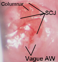
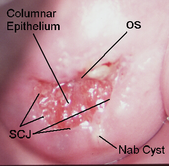
The columnar epithelium is a single-cell layer, mucous producing, tall epithelium that extends between the endometrium and the squamous epithelium. Columnar epithelium appears red and irregular with stromal papillae and clefts. With acetic acid application and magnification, columnar epithelium has a grape-like or "sea-anemone" appearance. It is found in the endocervix, surrounding the cervical OS, or (rarely) extending into the vagina.
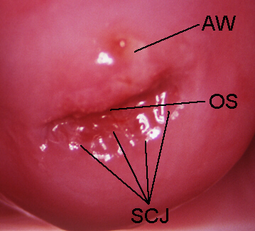
Generally, a clinically visible line seen on the ectocervix or within the distal canal (e.g., post-cryotherapy), which demarcates endocervical tissue from squamous (or squamous metaplastic tissue). This is an anatomical feature.
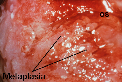
The physiologic, normal process whereby columnar epithelium matures into squamous epithelium. Squamous metaplasia typically occupies part of the transformation zone. At the squamocolumnar junction it appears as a "ghost white" or white-blue film with the application of acetic acid. It is usually sharply demarcated toward the cervical os and has very diffuse borders peripherally.
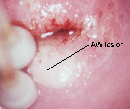
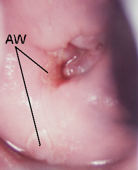
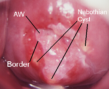 Acetowhite (AW). A transient, white-appearing epithelium following the
application of acetic acid. Areas of acetowhiteness correlate with higher
nuclear density.
-
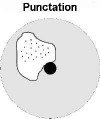
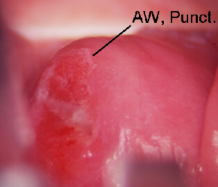
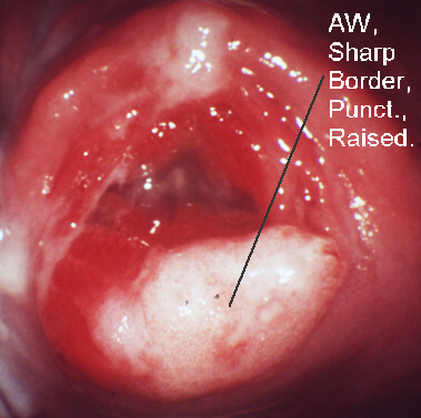
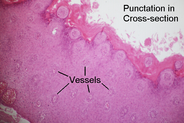 Punctation. A stippled appearance to capillaries seen end-on, often found
within acetowhite area appearing as fine to coarse red dots.
-
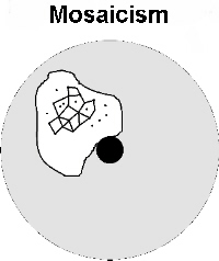 Mosaicism. An abnormal pattern of small blood vessels suggesting a confluence
of "tile" or "chickenwire" reddish borders.
Leukoplakia (hyperkeratosis). Typically an elevated, white plaque seen prior to the application of acetic acid.
Abnormal blood vessels. Atypical, irregular vessels with abrupt courses
and patterns, often appearing as commas, corkscrews, or spaghetti. No definite
pattern is recognized, as with punctation or mosaicism. Suspect invasive
cancer. Complex pattern consisting of roughened, irregular cervical epithelium,
typically with abundant irregular vessel patterns. Blood vessels take bizzare
forms, which appear as commas, hair pins, spaghetti, or long, dilated,
unbranching vessels with irregular diameters.
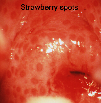
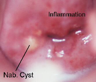 Cervicitis may cause abnormal Pap smears and make colposcopic assessment
more difficult. Many authorities recommend treatment before biopsy when
a STD is strongly suspected.
Less Severe > > More Severe
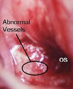 Atypical vessels usually indicate severe dysplasia or cancer. Acetowhite
areas that have sharp geographic borders and a dimension of thickness or
roughness are likely to be histologically more severe. Furthermore, all
other things being equal, the presence of vessel atypia in any lesion implies
more severe dysplasia. (5, 6)
A more formal system of assessing the severity of cervical dysplasia
is the Reid Colposcopic Index. (5, 6) It uses a point system to grade the
lesion margins, color, blood vessel pattern, and strong iodine staining
characteristics. It is more objective but not universally accepted as better
to classic subjective grading.
 Apply topical 20% benzocaine (Hurricane Solution) to decrease pain. It
is effective in 30 to 45 seconds. Perform an Endocervical Curettage (ECC)
before taking any biopsies unless the resultant blood would make biopsy
difficult. Use a Kevourkian curette (preferably without a basket) and scrape
the canal, 360 degrees, twice. The sample appears as a coagulum of mucus,
blood, and small tissue fragments. Use ring forceps or a cytobrush to gently
retrieve the sample. Submit on paper and label "ECC." Do not do an ECC
on pregnant patients.

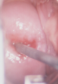
 Next, perform cervical biopsy. Biopsy posterior areas first to avoid blood
dripping over future biopsy sites. The cervix can be manipulated with a
Q-tip or hook if necessary to provide an adequate angle for biopsy.
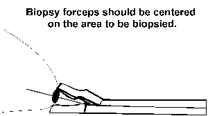
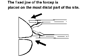 Align the forcep radially from the os so that the fixed jaw of the forcep
is placed on the most posterior part of the site. The jaws should be centered
over the area to be biopsied. Biopsies should be approximately 3 mm deep
and should include all areas with vessel atypism. It is not necessary to
include normal margins with biopsy samples. If bleeding is profuse from
a particular site and more biopsies are needed, apply a Q-tip to the area
and proceed with the next biopsy.
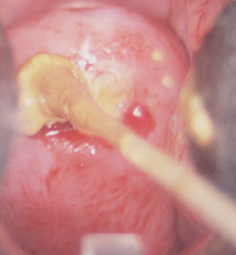 Do not apply Monsel's solution until all biopsies are completed. Apply
pressure first and Monsel's solution if needed to bleeding sites. The Monsel's
should be as thick as toothpaste to be most effective. Swab out the excess
Monsel+blood debris, that appears as a nasty black substance which eventually
will pass and may cause alarm (and potential late night phone calls).
Be concerned if a significant discrepancy is found between the colposcopic impression, Pap cytology, and biopsy histology. Be especially concerned if the biopsy reports are significantly less than Pap cytology. For instance, a Pap smear indicating carcinoma-in-situ and biopsies of only mild dysplasia could signify that the worst area was not biopsied. In general, a difference of one grade (i.e., Pap = CIN 2 and biopsy = CIN 3) is common and acceptable. Do not freeze any cervix until you have adequately and sufficiently explained any discrepancy between histology and cytology. If the discrepancy cannot be explained, conization is indicated. (2) Repeating colposcopy is forgivable, even in the hands of the best. Freezing invasive cancer is not.
Cone biopsy (cold cone, laser, or LEEP cone) is indicated if the endocervical curettage sample reveals dysplasia. It is a sin to freeze the cervix with disease in the canal. "Positive" ECC's are sometimes a result of contamination with dysplastic lesions at the verge of the os. Nonetheless, do not assume this!
Know your limitations! Never be afraid to call in help with an uncertain lesion or result.
 Follow-up after treatment is in 4- to 6-month intervals for 2 years, with
colposcopy or colposcopy intersperced with Pap smears. Recurrence is most
common in the first 2 years after therapy. Recurrences are most common
in the os and on the outside margins. A positive margin on a LEEP specimin
requires colposcopic follow-up.
2. McCord ML, Stovall TG, Summitt RL, Ling FW. Discrepancy of cervical cytology and colposcopic biopsy: Is cervical conization necessary? Obstet Gynecol 1991; 77:715-9.
3. Sadan O, Frohlich RP, Driscoll JA, Apostoleris A, Savage N, Zakust H. Is it safe to prescribe hormonal contraception and replacement therapy to patients with premalignant and malignant uterine cervices? Gynec Oncol 1986; 34:159-63.
4. Brotzman GL, Apgar BS. Cervical intraepithelial neoplasia: Current management options. J Fam Pract 1994; 39:271-8.
5. Reid R, Campion MJ. HPV-associated lesions of the cervix: Biology and colposcopic features. Clin Obstet Gynecol 1989; 32:157-79.
6. Reid R, Scalzi P. Genital warts and cervical cancer. VII. An improved colposcopic index for differentiating benign papillomaviral infections from high-grade cervical intraepithelial neoplasia. Am J Obstet Gynecol 1985; 153:611-8.
7. Stafl A, Wilbanks GD. An international terminology of colposcopy:
Report of the nomenclature committee of the International Federation of
Cervical Pathology and Colposcopy. Obstet Gynecol 1991; 77:313-4.