|
This pamphlet will explain:
- Why hysteroscopy may be used
- How it is done
- What to expect before and after the procedure
Uses of Hysteroscopy
|
Hysteroscopy may be used for diagnosis, treatment or
both. |
Hysteroscopy is minor surgery that may be done in a doctor's
office or operating room with local, regional, or general
anesthesia. In some cases, little or no anesthesia is needed. The
procedure poses little risk for most women. Hysteroscopy may be used
for diagnosis, treatment or both.
Diagnostic Hysteroscopy
Hysteroscopy can be used to diagnose some problems in the uterus.
It also can be used to confirm the results of other tests, such as
hysterosalpingography (HSG).
The hysteroscope is sometimes used with other instruments or
techniques. For instance, it may be done before dilation and
curettage (D&C) or at the same time as laparoscopy. In a
D&C, the cervix is widened (dilation) and part of the lining of
the uterus is removed (curettage). In laparoscopy, a slender,
telescope-like device is inserted into the abdomen through a tiny
incision (cut) made through or just below the navel. Hysteroscopy
also may be used for other conditions.
Abnormal Uterine Bleeding. A woman has this condition if
she has heavier or longer periods than usual, bleeds between
periods, or has any bleeding after her periods have stopped at
menopause. Hysteroscopy may help the doctor find the cause of
abnormal bleeding that other methods have not found. It may be used
to take a biopsy.
Infertility. A couple may not be able to achieve pregnancy
for a number of reasons. Sometimes the cause of female infertility
is related to a defect in the shape or size of the uterus. One
example of this is a septate uterus (a thin sheet of tissue divides
the inside of the uterus into two sections). Hysteroscopy may find
these problems if other tests do not.
|
 |
|
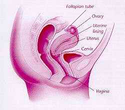
The uterus is a muscular organ located in the pelvis.
It is broad at the top and narrow at the bottom. At each side
of the upper part, a fallopian tube leads outward toward an
ovary. The ovaries contain many eggs, or ova, and normally
release one during each menstrual cycle. The tubes carry a
fertilized egg from the ovaries to the uterus. The lower end
of the uterus, called the cervix, is a narrow channel with a
small opening. It opens into the
vagina. |
Repeated Miscarriages. Some women, although able to get
pregnant, lose the fetus to miscarriage - the loss of a pregnancy
before 20 weeks. Hysteroscopy can be used with other tests to help
find the causes of repeated miscarriage.
Adhesions. Bands of scar tissue, or adhesions, may form
inside the uterus. This is called Asherman syndrome. These adhesions
may cause infertility and changes in menstrual flow. Hysteroscopy
can help locate adhesions.
Abnormal Growths. Sometimes benign growths, such as polyps
and fibroids, can be diagnosed with the hysteroscope. Hysteroscopy
might help a doctor to biopsy a growth in the uterus to find out
whether it may be cancer or may become cancer.
Displaced IUDs. An intrauterine device (IUD) is a small
plastic device inserted in the uterus to prevent pregnancy. In some
cases, it moves out of its proper position inside the uterus. It
then embeds itself in the uterine wall or the tissue around it.
Sometimes hysteroscopy can be used to locate an IUD.
|
 |
|
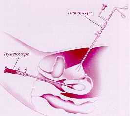
Laparoscopy sometimes is done along with
hysteroscopy. |
Operative Hysteroscopy
When hysteroscopy is used to diagnose certain conditions, it may
be used to correct them as well. For instance, uterine adhesions or
fibroids often can be removed through the hysteroscope. Sometimes
hysteroscopy can be used instead of open abdominal surgery. Often it
will be done in an operating room with general anesthesia.
The hysteroscope is used to perform endometrial ablation - a
procedure in which the lining of the uterus is destroyed to treat
some causes of heavy bleeding. After this is done, a woman can no
longer have children. For this procedure, the hysteroscope is
sometimes used with other instruments, such as a laser or a
resectoscope. The resectoscope is a specially designed telescope
with a wire loop or a rollerball at the end. Using electric current,
any of these tips can be used to destroy the uterine lining.
Endometrial ablation is done in an outpatient setting in most cases.
|
 |
|
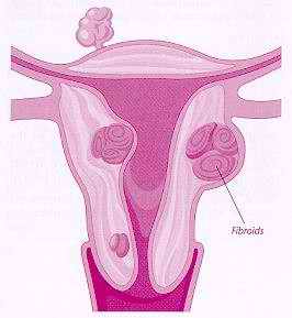
A fibroid is a benign growth that may form inside the
uterus. |
What to Expect
Hysteroscopy is a safe procedure. Problems such as injury to the
cervix or the uterus, infection, heavy bleeding, or side effects of
the anesthesia occur in less than 1 percent of cases.
Before Surgery
Hysteroscopy is best done during the
first week or so after a menstrual period. This allows a better view
of the inside of the uterus.
If you are having a hysteroscopy in a hospital, you may be asked
not to eat or drink for a certain time before the procedure. Some
routine lab tests may be done. You will be asked to empty your
bladder. Then your vaginal area will be cleansed with an antiseptic.
Anesthesia
Hysteroscopy may be performed with local,
regional, or general anesthesia. The type used depends on a number
of factors. This includes whether other procedures are being done at
the same time. Where you have your surgery - in your doctor's office
or in the hospital - also may affect the kind of pain relief used.
You will want to discuss your options with your doctor.
Before the procedure, your doctor may prescribe a medication to
help you relax (a sedative) before the anesthetic is injected. When
a local anesthetic is used, it is injected around the cervix to numb
it. You will remain awake during the procedure. You may feel some
cramping.
With regional anesthesia, a drug will be injected to block the
nerves that receive feeling from the pelvic region. You will be
awake but will not feel any pain. The anesthetic will be given
through a needle or tube in your lower back. This is called a spinal
or epidural.
With general anesthesia, you breathe a mixture of gases through a
mask. You will not be conscious during the surgery. After the
anesthetic takes effect, a tube may be put down your throat to help
you breathe.
The Procedure
Before a hysteroscopy, the opening of
your cervix may need to be dilated (made wider) with a special
device. The hysteroscope then is inserted through the vagina and
cervix and into the uterus.
A liquid or gas may be released through the hysteroscope to
expand the uterus so that the inside can be seen better. A light
shone through the device allows the doctor to view the inside of the
uterus and the openings of the fallopian tubes into the uterine
cavity. If surgery is to be done, small instruments will be inserted
through the hysteroscope.
For more complicated procedures, a laparoscope may be used at the
same time to view the outside of the uterus. In this case, a gas is
flowed into the abdomen. The gas expands the abdomen. This creates a
space inside by raising the wall of the abdomen and moving it away
from the internal organs. This makes the organs easier to see. Most
of this gas is removed at the end of the procedure. This procedure
is not done in the office.
Recovery
If local anesthesia was used, you will be able
to go home in a short time. If regional or general anesthesia was
used, you may need to be watched for some time before you go home.
You may feel a pain in your shoulders if laparoscopy was done
with hysteroscopy or if gas was used during hysteroscopy to inflate
the uterus. In most cases, the pain passes quickly as the gas is
absorbed. You may feel faint or sick or you may have slight vaginal
bleeding and cramps for a day or two. Get in touch with your doctor
if you have:
- A fever
- Severe abdominal pain
- Heavy vaginal bleeding or discharge
Finally...
Because hysteroscopy allows your doctor to see the inside of the
uterus, it may permit a diagnosis of some medical problems. It also
may be used to treat them. The procedure and recovery time are brief
in most cases.
Glossary
Adhesions: Scars that bind together affected surfaces of
the tissues inside the abdomen or uterus.
Anesthesia: Relief of pain by loss of sensation.
Biopsy: A minor surgical procedure to remove a small piece
of tissue that is then examined under a microscope in a laboratory.
Cervix: The lower, narrow end of the uterus, which
protrudes into the vagina.
Fibroids: Benign (noncancerous) growths that form on the
inside of the uterus, on its outer surface, or within the uterine
wall itself.
Hysterosalpingography (HSG): A special X-ray procedure in
which a small amount of fluid is injected into the uterus and
fallopian tubes to detect abnormal changes in their size and shape
or to determine whether the tubes are blocked.
Menopause: The process in a woman's life when ovaries stop
functioning and menstruation stops.
Polyps: Benign (noncancerous) growths that develop from
membrane tissue, such as that lining the inside of the uterus.
Uterus: A muscular organ located in the female pelvis that
contains and nourishes the developing fetus during pregnancy.
Vagina: A passageway surrounded by muscles leading from
the uterus to the outside of the body, also known as the birth
canal.
|


